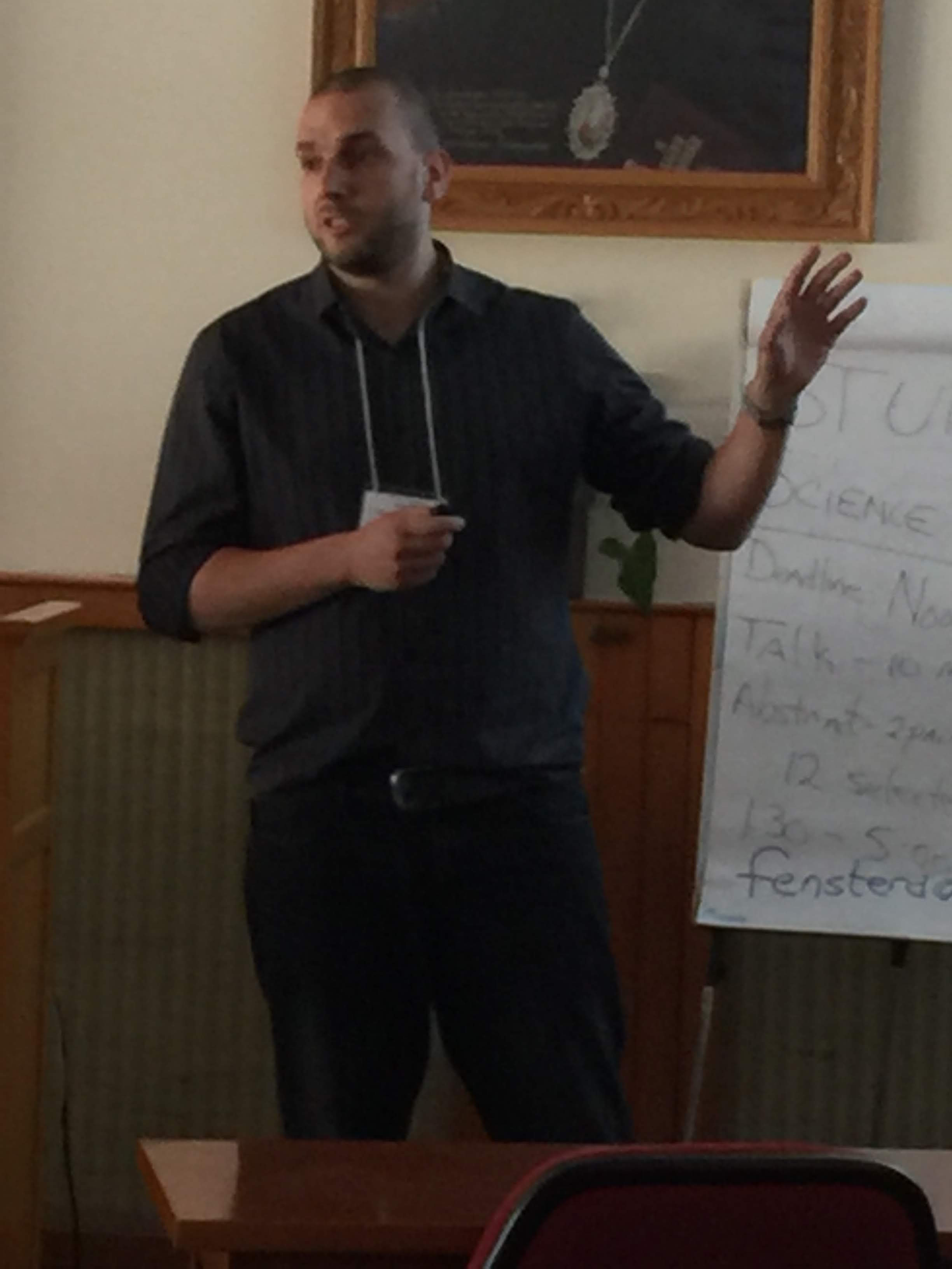
Péter Maróti
|
Phoenix Orthoses-SMA (smart memory alloy) and additive manufacturing technologies in active upper-limb exoskeleton development
Despite the effective progress of medical care in stroke patients, it's still the second leading cause of death in worldwide. In the US, stroke is the main cause of long-term severe adult disability, and according to the recent epidemiologic reports the number of young adults affected by stroke is escalating. The outcome of stroke has enhanced recently, but the consequences are still devastating due to the long term complications affecting the mobility, psychological status and loss of functional independency for everyday life. A new perspective for the motor rehabilitation is the integration of the robotic therapy (RT) to the conventional physiotherapy (CT). RT studies have shown that, combined with conventional therapy, it is more successful than classical methods. Active orthoses or exoskeletons are a dynamically evolving, interdisciplinary research field; therefore, there are several promising projects all over the world, aiming to create the ideal device. The users' needs and expectations are key factors in device development, which means user-friendly, cost-effective, aesthetic solutions are favoured, which gives the maximum support in case of AoDLs (activities of daily living). Also, decreasing the noise of operation is a key element and important aim from the user's perspective. Reflecting the mentioned need for SMAs (shape memory alloys) as an active part of the device can be a great solution. NiTinol has important SME (shape memory effect) and SE (superelasticity) characteristics. Combined with additive manufacturing (AM) technologies, the cost effectivitness can be enhanced. Our aim was to develop a device, with a complete production workflow plan, which fulfills the observed patient-needs, could be personalized and provides a fast, cost effective solution for post-stroke patients. The device is aimed to help the rehabilitation of the upper limb (arm), targeting the grasping movement.
|
| |
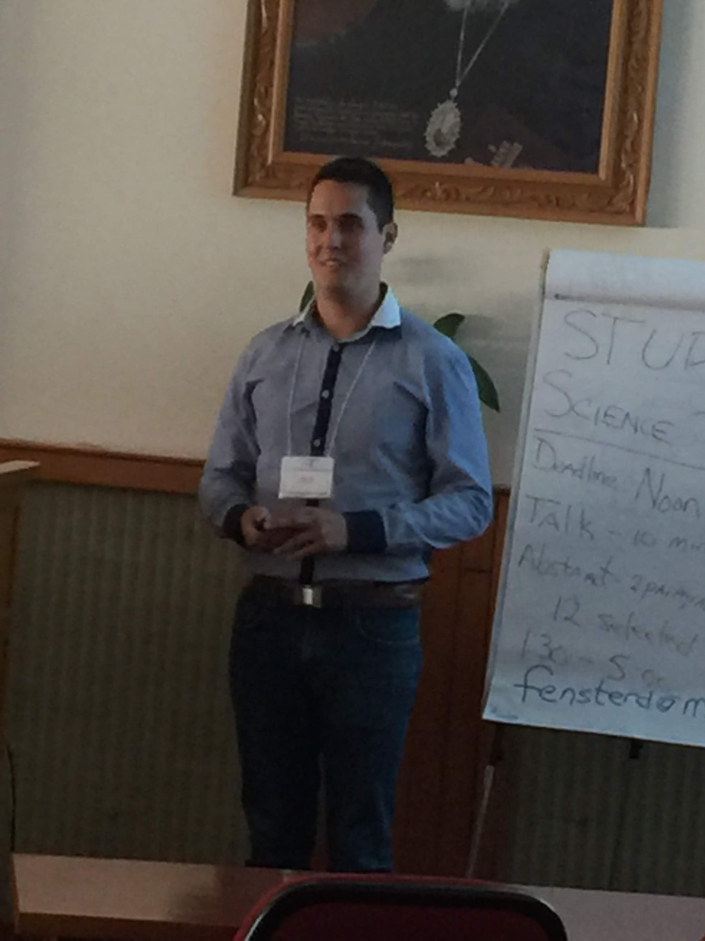
Péter Varga
|
Precision Medicine in Cardiac Surgery: Application of 3D Printed Surgical Guides in Left Ventricle Aneurysm – a pilot study
Introduction: Left ventricular aneurysm is defined as circumscribed, thin-walled, non-contractile out-pouching of the ventricle that occur as a result of myocardial infarction, causing impaired left ventricular function, arrhythmia and valvular dysfunction. Several surgical methods are used for ventricular restoration of aneurysms, which are all excessively dependent upon creating the advantageous left ventricular morphology and function. Although there is no golden standard technique to be applied -as every aneurysm morphology is different-, so a personal medical imaging data-based precision medicine solution can be beneficial in the outcome.
Materials and Methods: Surgical guides are advantageously applied in the perioperative planning and implementation of interventional and surgical procedures. However, thus far, these applications were mostly confined to planning within a static environment. In consideration of the dynamic changes in tissues, such as myocardium, in which the functional outcome mostly is dependent upon the effective approach to strategic planning, notably, is lacking in experience and in the consideration of the most suitable method. Pathological models were segmented (3D Slicer 4.7, Slicer Inc.) from personal DICOM data. Finite element analyses (Ansys 19.0, Ansys Inc.) were run to simulate the most advantageous postoperative left ventricle morphology. A fused deposition modelling 3D printer (Craftbot 2.0., Craftunique Ltd.) was utilized with polylactic acid filament (Philamania Ltd.) to create the models and the surgical guides. Gamma sterilization was performed (Dispomedicor Inc.) on the devices that were brought to the surgical theatre.
Results: In the framework of this pilot study a patient-specific model of the heart and tactical guides were created in support of effective patch sizing and orientation. It was based on personal medical imaging data using computational fluid dynamics analysis and virtual planning. The 6th week post-operative results showed a 48% improvement in left ventricle function (ejection fraction from 25% to 37%), mitral regurgitation decreased from 49ml to 18 ml, left ventricle end-systolic volume index decreased from 95ml/m2 to 63ml/m2 and no mitral valve replacement was needed.
Conclusion: Applying a multi-modality virtual modelling and simulation method can assure precision and accuracy, with respect to effectively planning both the left ventricular and valvular beneficial functional outcome.
|
| |
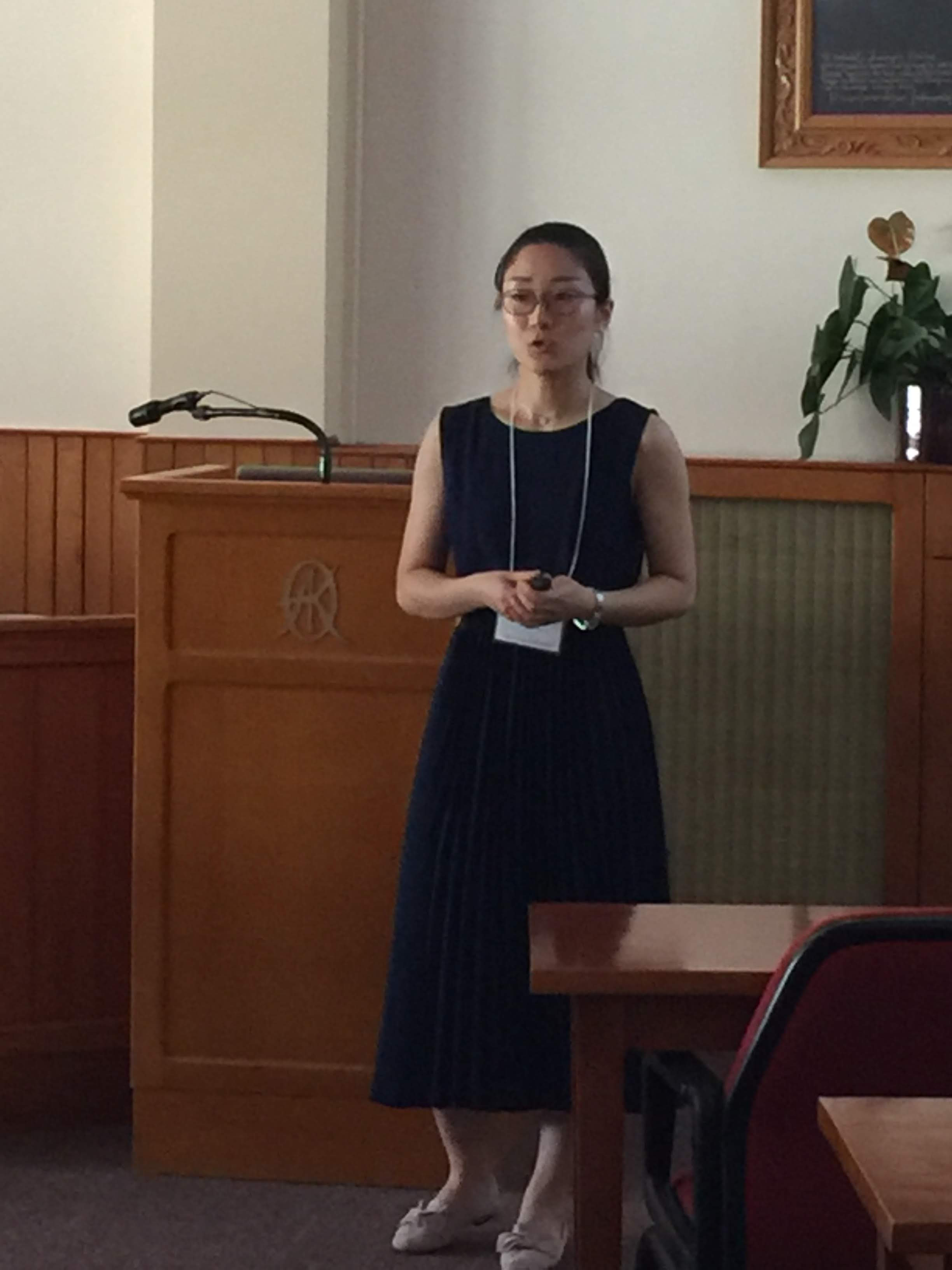
Haoran Ren
|
BIO-X Summer School Presentation by Haoran Ren
Functional near-infrared spectroscopy (fNIRS) is a potential non-invasive monitoring technique for evaluating functional activity of the cerebral cortex by investigating the cerebrovascular hemodynamic changes. The two length of near-infrared light is send into the tissue and then the diffusing light is measured by the optodes, which can estimate the cerebrovascular hemoglobin concentrations. The changes of the concentrations of oxygenated and deoxygenated hemoglobin detected by the NIRS reflect the brain activity, which is based on the mechanism of neurovascular coupling. fNIRS has the advantages of low-cost, portability and robust to motion artifacts compared with the other neuroimaging modalities including functional magnetic resonance imaging (fMRI) and positron emission tomography (PET). Considering the advantages, fNIRS has been applied in various fields, such as neurology, psychiatry, psychology/education and basic research, especially in clinical setting or physical activity scenarios, even for neonates monitoring.
Human brain is a complex biological system which has many complex regulatory mechanisms. The complexity analysis provides an effective way to quantify the changes of brain activity. The modified multiscale entropy (MMSE) algorithm was employed to assess the complexity of fNIRS signals which may reflect the changes of brain activity when people underwent brain injury. The results that the mean MMSE of oxyhemoglobin values was lower in TBI patients compared to healthy subjects, indicated that MMSE was feasible to measure complexity of cerebral near-infrared spectroscopy signals in TBI patients, and that brain injury was associated with the decreased complexity of cerebrovascular reactivity. Moreover, measurement of complexity of brain signals has potential to provide significant guidance for rehabilitation.
|
| |

Isle Bastille
|
BIO-X Summer School Presentation by Isle Bastille
The objectives of this talk are threefold:
- Exposing the audience to the basic biology of the blood-brain barrier (BBB)
- Highlighting the gap in knowledge between the basic cellular biology of the BBB and current therapeutic drug development strategies
- Introducing a novel hypothesis of the sub-cellular trafficking mechanisms that prevent drug delivery to the brain
The central nervous system (CNS) requires a tightly regulated microenvironment for proper neuronal function. To accomplish this, specialized endothelial cells which line the walls of cerebral blood vessels, comprise the blood-brain barrier (BBB). The BBB serves to restrict passage of substances between the blood and surrounding CNS tissue. BBB breakdown is implicated in various neurological diseases. At the same time, the intact BBB prevents the delivery of intravenously administered therapeutic drugs to the surrounding CNS tissue. In fact, it is estimated that the BBB prevents over 95% of therapeutic drugs, including large molecules such as antibodies from accessing the brain. It is therefore crucial to thoroughly understand the basic cell biology of the BBB to efficiently treat neurological disease.
In this talk, I will present the current approaches to delivering therapeutics across BBB endothelial cells to the brain. Currently, most therapeutic drug development and delivery strategies are focused on co-opting intact endothelial cell endocytic mechanisms. These strategies operate on the assumption that molecules that are endocytosed into BBB endothelial cells are subsequently transcytosed and released to the surrounding CNS tissue. However, the sparsity of successful CNS therapeutics indicates a fundamental disconnect between pharmaceutical drug development and a thorough understanding of BBB basic cellular biology.
My PhD work is focused on understanding BBB endothelial cell trafficking mechanisms. Currently, I propose that most molecules that are endocytosed into BBB endothelial cells are quickly trafficked to the lysosomal system to be degraded. In fact, I hypothesize that lysosomal degradation in BBB endothelial cells is a major bottleneck to efficient drug delivery. However, this basic cellular process is rarely considered in current therapeutic drug design and BBB studies. I hope that this presentation and my future work will provide needed insight into BBB cellular biology and spark novel approaches in therapeutic drug development for the brain.
|
| |
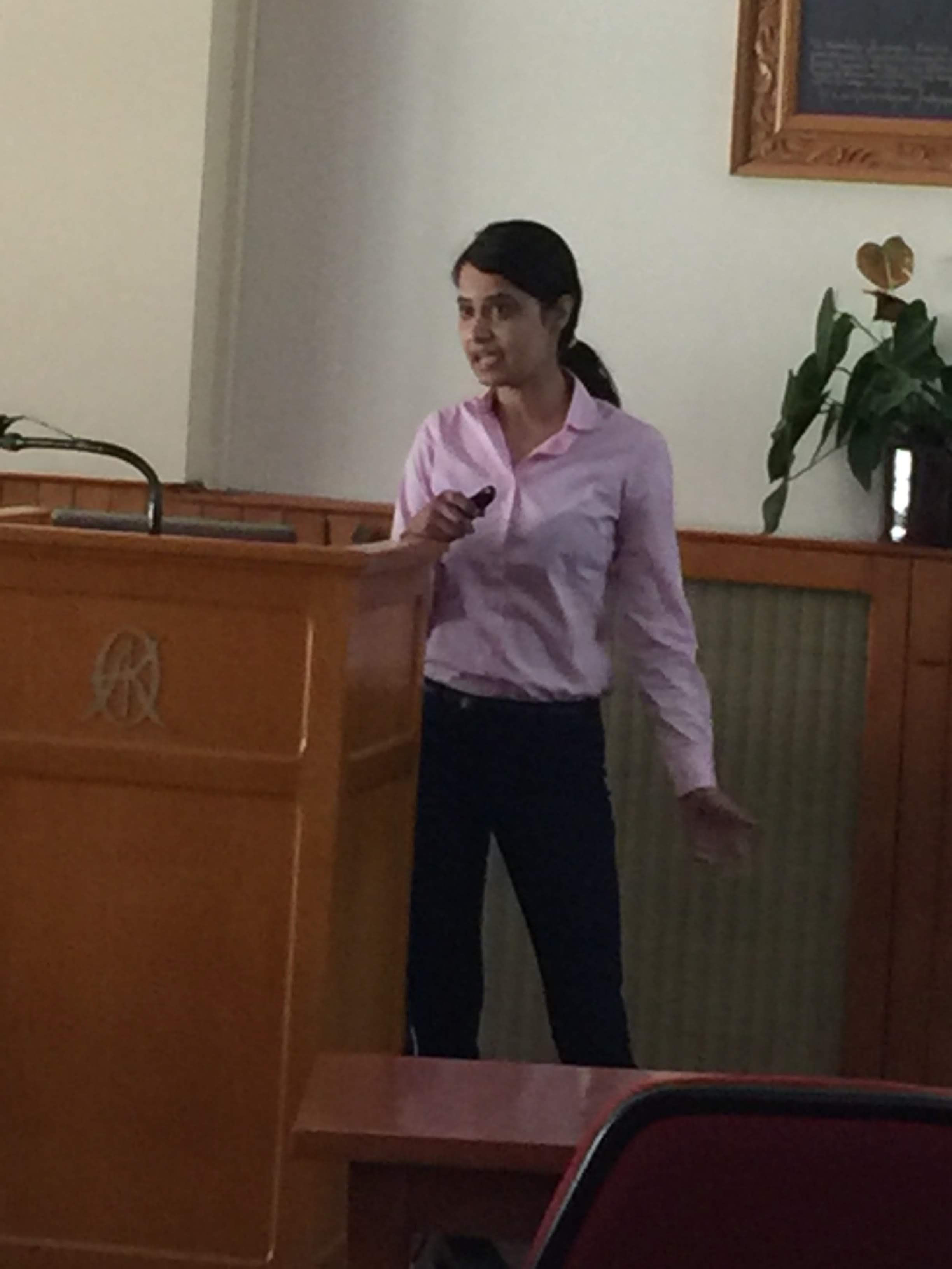
Ruta Deshpande
|
Exploring cortical evoked potentials as a biomarker to improve Deep Brain Stimulation therapy
DBS therapy is a treatment for severe childhood-onset dystonia that usually takes weeks or months to have its full effect. The therapy, which involves brain surgery to implant a 'pacemaker' for the brain, can have a profound impact on dystonic symptoms for some individuals, yet for others the changes are subtler.
We are measuring brain activity in relation to DBS (using scalp EEG) to get a better understanding of how the therapy interacts with the brain and to optimize stimulation parameters for a given child. In addition, we are performing recordings during the implant surgery to record signals from brain areas that are otherwise inaccessible. Such recordings will help us get a deeper understanding of abnormalities and dystonia and may help improve DBS therapy.
I have helped to develop computational models in MATLAB to understand the physiological mechanisms of Deep Brain Stimulation and get a better understanding of interaction of the brain with therapy. The basic steps include averaging, squaring and thresholding of the signal.
We identify the best target with respect to the response seen in the post-operative EEGs, which helps optimize stimulation parameters for all patients. These EEG cortical evoked potentials can be used as a biomarker to make closed loop stimulation a reality.
|
| |
Renee Keller
|
Investigating the Effect of Perinatal Nicotine Exposure on Dopaminergic Neurons in the VTA using miRNA Expression Profiles
Maternal smoking during pregnancy is associated with developmental, cognitive, and behavioral disorders, including low birth weight, attention deficit hyperactivity disorder, learning disabilities, and drug abuse later in life. Nicotine, the primary addictive component in tobacco, has been shown to modulate changes in gene expression when exposure occurs during neurodevelopment. Dopaminergic (DA) neurons originating from the ventral tegmental area (VTA) of the brain, which are stimulated by nicotine and other stimuli, are widely implicated in the natural reward pathway that is known to contribute to addiction. In recent years, microRNAs have been implicated in disrupting regulatory mechanisms due to their capability of targeting multiple genes and thus inducing downstream effects along many pathways. In order to investigate miRNA expression of dopaminergic neurons from the VTA, we employed patch clamping to identify and harvest both DA and non-DA neurons from the brain of rats exposed to nicotine during gestation for use in single-cell RT-qPCR. Our data indicated that miR-140-5p and miR-140-3p were upregulated in DA neurons; while miR-140-3p and miR-212 were differentially expressed in non-DA neurons. A functional enrichment analysis was also performed on our miRNA-gene prediction network.
|
| |
Dhruv Seshadri
|
Translating the Clinical Utility of Commercial Wearable Sensors for Medical Applications title
Over the last decade, there has been an expansion and development of wearable technology within the field of sports medicine to monitor human performance. The recent technology drive has enabled the application of biomechanical and biochemical monitoring of athletes in real time to help guide clinical decision-making via the translation of the acquired data into actionable protocols for team physicians and athletic trainers. Quantitative analysis of biometric sensor data has shown promise in the field of sports medicine to drive patient specific recovery and rehabilitation protocols to maximize performance while minimizing the risk of injury. From our perspective, over-engineering of biomedical sensors has drastically limited their clinical utility. Thus, to circumvent such inefficiencies and develop technologies to complete the bench to bedside paradigm, we seek to assess the utility of this field and identify critical path elements to enable such translation. The objective of our team-centered work is two-fold: 1) build collaborative relationships with key opinion leaders in the medical field and 2) develop testable hypotheses leveraging emerging IOT technology utilizing our expertise in materials science, big data, and engineering . Work disseminated from this abstract has resulted in the emergence of projects reflecting our team-centered approach utilizing wearable sensors for sports medicine, cardiology, and emergency medicine.
Our inter-disciplinary team of engineers, orthopedic surgeons, anesthesiologists and cardiologists, enables the ability to produce ideas and outcomes with direct added value to the medical field. To date, our collaborators utilized wearable devices to assess the effect of player load on soft tissue injuries in professional athletes. Athletes were studied over the course of two years and were found to be at a higher risk of injury during rapid changes to player load as compared to average load over a one-week timeframe. Future work is needed to utilize sensor data to establish direct protocols to mitigate the risk of injury among athletes and improve athlete performance. In this talk, I will highlight our collaborative work in the sports medicine field, discuss current work going on with colleagues at the University of Grenoble Alpes in developing conductive biodegradable materials and epidermal electrodes, and discuss the utilization of analytical platforms such as Natural Language Processing (NLP) to enhance clinical decision making.
|
| |
Mackenzie Grubbb
|
BIO-X Summer School Presentation by Mackenzie Grubb
The use of magnetic nanoparticles for the delivery of genetic material into the cell has been proven advantageous through a number of different techniques and in a wide variety of cell types. This application of magnetic particles in gene transfection, termed magnetofection, has been shown to improve the transfection efficiency, decrease the time needed for uptake, and increase the overall protein production. Further, when an oscillating magnetic array is applied the increased energy of the system has been shown to increase the uptake of the functionalized particles through endocytosis which again decreases the sedimentation time needed for transfection, as well as increase the overall transfection efficiency.
We have then taken this concept of the use of an oscillating magnetic array in magnetofection to be applied in different time points in the post-transfection period to test how this method may also be used to increase endosomal escape. We tested this method against a static magnetic array pre- and post-transfection. The cells that had been oscillated post-transfection at multiple different time points each showed varying degrees of sustained expression of the reporter vector for significant time over the static array post-transfection.
This technique is applicable over a variety of cell types and has the potential to impact many gene therapy techniques in vitro such as the biopharmaceutical industry for applications such as monoclonal antibody production.
|
| |

Mahum Siddiqui (left) and Nicole Philip (right)
|
Effect of LIV conditioned macrophages on pre-adipocytes for inflammatory regulation in diabetic patients
Under Dr. Stefan Judex of the Integrative Skeletal Adaptation and Genetics Lab at Stony Brook University, the main purpose of our study is the effect of LIV on macrophages and subsequently, pre-adipocytes. At high frequency oscillations, low intensity vibrations are known to have a pro-healing effect through increased expression of growth factors and a decreased expression of pro-inflammatory cytokines by macrophages. Though many studies have been done on the effect of LIV on wound healing, this study was geared towards its potential benefits on fat cells. We studied the altered gene expression of pre-adipocytes after treatment of LIV conditioned macrophage media.
Adipose cells and macrophages display a close relationship between inflammation factors and adiponectin production. Type 2 diabetic patients are known to show high inflammatory levels as well as decreased differentiation capacity of pre-adipocyte cells. These effects are a cause of chronic inflammation which ultimately affect the ability to lose weight and the susceptibility to other chronic issues. In order to study the potential benefits of LIV on diabetic patients, we exposed murine pre-adipocytes to media from high frequency/low-intensity vibration treated M0 and M1 activated macrophages. Data was collected on the gene expression of four proinflammatory cytokines: MCP-1, TNF-α, IL-6, and IL-1β, which are commonly produced at high rates in diabetic patients, as well as two anti-inflammatory genes, IFN-γ and IL-10. We are still in the process of collecting experimental data, but trials were carried out using a total of 24 samples over the course of four days with three days of vibration treatment, and one day of pre-adipocyte exposure.
|
| |
Jessica Eager
|
Macrophage Characterization of Diabetic Foot Ulcers
Dysfunctional wound healing is a major complication of both type 1 and type 2 diabetes. Diabetic foot ulcers (DFUs) occur in 15% of diabetic patients, leading to over 82,000 lower extremity amputations annually in the United States. The greatest challenge in treating DFUs lies within patient-to-patient variability: it is unknown why some patients respond to certain treatments, while others do not. The mechanisms behind impaired DFU healing are complex, although dysregulated inflammation and innate immune cell behavior, specifically macrophages, are strongly implicated. In normal wound healing, changes in macrophage activation states are essential for successful healing because they regulate the behavior of other important cell types. Over time, macrophages transition from a pro-inflammatory phenotype (M1) to a more anti-inflammatory phenotype (M2), which is believed to facilitate resolution of the healing process. The activities of both phenotypes, in the proper order, are required for successful healing. Deficient M1 activation at early stages of healing and/or inadequate M2 activation at later stages result in improper healing.
To assess this paradigm, our lab previously collected debrided tissue from 10 DFU patients and found that the ratio of M1 to M2 genes decreased over time for all DFUs that would ultimately heal within 12 weeks, whereas this ratio increased for those that did not close within that time frame (non-responders). These results suggest that non-responders to the standard of care may be optimal candidates for treatment with anti-inflammatory treatments, which would decrease the M1/M2a ratio of their tissue. Conversely, patients with low levels of M1 activation are expected to be non-responders to anti-inflammatory treatments because they would exacerbate their hypo-inflammatory state and inhibit the sequential M1-to-M2a activation. To these hypotheses, debrided DFU tissue was collected from ~40 subjects treated with either the standard of care or an anti-inflammatory treatment. After 12 weeks, subjects were classified as responders or non-responders based on whether their DFUs completely closed during this time period. DFU tissue samples collected from each group were analyzed for a panel of 175 genes that we have previously identified as markers of macrophage phenotypes. Work is ongoing to analyze the differences in gene expression between responders and non-responders to the standard of care as well as advanced care treatments in order to develop novel diagnostics with a precision medicine approach.
|
| |
Konstadina Kourou
|
Modeling biological data using dynamic bayesian networks for oral squamous cell carcinoma classification
We propose a computational approach for modeling the progression of Oral Squamous Cell Carcinoma (OSCC) through Dynamic Bayesian Network (DBN) models. RNA-Seq transcriptomics data, available from public functional genomics data repositories, are exploited to find genes related to disease progression (i.e. recurrence or no recurrence). Our primary aim is to perform a complete upstream analysis based on the differentially expressed genes identified. More specifically, a search for putative transcription factor binding sites (TFBSs), in the promoters of the input gene set, as well as an analysis of the pathways of the suggested transcription factors is conducted. Activities of transcription factors which are regulated by upstream signaling cascades are further discovered. These converge in certain nodes, representing molecules which are potential regulators of OSCC progression. The resulting gene list is further exploited for the inference of their causal relationships and for disease classification in terms of DBN models. The structure and the parameters of the models are defined subsequently, revealing the patient status during the follow-up period after surgery.
The objectives of the proposed methodology are to: (i) accurately estimate OSCC progression, and (ii) provide better insights into the regulatory mechanisms of the disease. Moreover, we can conjecture about the interactions among genes based on the inferred network models. The proposed approach implies that the resulting regulatory molecules along with the differentially expressed genes extracted, can be considered as new targets, and are candidates for further experimental and in silico validations.
|
| |

Amanda Nash
|
Characterizing the Anti-inflammatory Microevironment of Mesenchymal Stem Cells
Mesenchymal stem cells (MSCs) are of great interest to the scientific community due to their powerful differentiation capabilities and the immunosuppressive properties that allow them to survive host immune rejection. We have previously demonstrated that human umbilical cord stem cells (hUMSCs) secrete a specialized extracellular matrix or glycocalyx which is capable of suppressing the inflammatory response. The anti-inflammatory properties of the components of this glycocalyx allowed the UMSC from humans to survive xenograft rejection when transplanted into immunocompetent mice. For this project we are investigating the specific mechanism by which these UMSC suppress the host immune response.
In addition to attempting to elucidate the mechanism of immune response suppression by the UMSCs, we are also taking strides to increase the immunosuppressive properties of other types of mesenchymal stem cells, such as bone marrow stem cells (BMSCs). For this study, we are manipulating the components of the extracellular matrix of the BMSCs to mimic the extracellular environment of the UMSCs. Because we believe that the extracellular environment of the UMSC is essential to the anti-inflammatory properties of the cells, we believe that reverse engineering of the BMSC extracellular environment will lead to an increase in their ability to exhibit anti-inflammatory properties. The specific components we focus on in this project include 1) hyaluronan (HA), 2) heavy chains (HCs), 3) tumor necrosis factor (TNF)-stimulating gene 6 (TSG-6). The success of this project could lead to a decrease in the percent likelihood of having bone marrow transplant rejections due to activation of the host immune response in the future.
|
| |
Krisztina Bánfai
|
Significance of Wnt4-exosomes in thymic senescence
It is well-known, that Wnt4 plays an essential role during the development of the thymus via regulating thymopoiesis and maintaining the cellular composition of TECs (thymic epithelial cells). When thymic senescence starts Wnt4 is down-regulated, while PPARγ is up-regulated and triggers adipose involution. Of note, miR27b was described to suppress PPARγ. Both the Wnts and various miRNA species have been reported to reside in exosomes in other contexts. Our aim was to confirm the presence of Wnt4 and miR27b in TEC exosomes, detect their effect and follow their migration both in vitro and in vivo.
Exosomes were collected from control and Wnt4 over-expressing TECs. Transmission electron microscopy was used to visualize exosomes. Exosomal miR27b levels were measured by TaqMan qPCR, while Wnt4 protein content was assessed by ELISA. DiI-labeled exosomes were injected into mice intravenously to detect homing in the body. Using transmission electron microscopy exosomes were detected in the range of 50-100 nm as expected. TaqMan miRNA assay measured elevated miR27b levels, while ELISA showed high Wnt4-content in Wnt4- exosomes compared to control exosomes. To create a cellular aging model, steroid (Dx)- induced TECs were used. Dx accelerated aging, but Wnt4-containing exosomes could efficiently counteract Dx-induced senescence. Finally, in vivo injected DiI-labeled Wnt4- exosomes showed detectable homing to the thymus. According to our results Wnt4 and miR27b are both present in TEC exosomes. Our findings indicate that Wnt4 may act as an inhibitor of thymic involution via miR27b. However, further studies are required.
|


